


Coral Reef Biology
A coral reef is built by polyps a few millimetres across that live with algae inside their cells and lay down aragonite beneath them. This unit explains the partnership that lets a reef thrive in nutrient-poor water, the chemistry and the speed of reef growth, Darwin's explanation of atolls and the drill hole that confirmed it, the grazers that keep a reef clean, and what happens inside a coral cell when the sea becomes too warm. It ends with NOAA's Degree Heating Weeks, the four global bleaching events, and the corals of American Samoa that tolerate heat better than their neighbours.
A coral reef is the largest structure built by living things, large enough to be seen from orbit, and it stands in some of the clearest water of the ocean, water that is also among the poorest in nutrients. The builders are polyps a few millimetres across, each one housing algal cells that feed it, and together they have raised limestone platforms more than a kilometre thick and a Great Barrier Reef 2,300 kilometres long. This unit explains how the partnership works, how the limestone is made, why Darwin could explain an atoll without seeing beneath one, what lives on a reef and keeps it clean, and what happens inside a coral cell when the sea becomes too warm. It ends with the corals that tolerate heat better than their neighbours, which are the reason the story is not yet over.
1 The coral animal and its partner
1.1 Polyp, colony, and skeleton
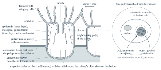
A reef-building coral is a colony of cnidarian polyps of the kind Unit 4 described, with two additions that change everything, a skeleton of limestone beneath the animal, which this subsection describes, and algae inside its cells, which the next one describes. Each polyp sits in a cup of its own making, and the cups together form a skeleton of calcium carbonate that the living tissue covers like a skin. Figure 1 shows one polyp cut lengthwise. The animal has the two cell layers of every cnidarian, an outer epidermis and an inner gastrodermis, with the jelly-like mesoglea between them, a mouth that leads through a short pharynx into the gastrovascular cavity (the single internal cavity that serves the polyp as a gut), and a ring of tentacles armed with stinging cells. In most reef builders the polyp is 1 to 3 millimetres across, about the size of a grain of rice, and a colony the size of a table holds hundreds of thousands of them. The polyps are joined by a sheet of tissue called the coenosarc, which runs over the skeleton between the cups, and food caught by one polyp is shared with its neighbours through the connected cavities.
The cup, called the corallite, is made of aragonite, which is one of the two crystal forms of calcium carbonate (\(\mathrm{CaCO_3}\)). Aragonite is denser than calcite, which is the form found in the shells of most molluscs and in the plates of the coccolithophores of Unit 3, and it also dissolves more readily than calcite does; this difference matters in Section 2. Radial plates called septa rise from the floor of each cup and give the skeleton its strength and each species its pattern. The cells that build all of this form a thin layer at the base of the polyp, the calicoblastic layer, which lies directly on the skeleton and secretes it from below. The polyp therefore lives on top of its own house and keeps adding to the floor, so that the colony grows upward and outward and the older skeleton below is left empty.
The stony corals form the order Scleractinia of the class Anthozoa, with about 1,600 described living species, of which roughly 800 live with symbiotic algae and build reefs; the others, among them the cold-water corals of Section 6, live without symbionts and often in the dark. Stony corals appear in the fossil record in the Triassic period, about 240 million years ago, and the colonial species grow as branching staghorns, flat tables, solid domes (called massive corals, a name for their build rather than their size), and thin sheets that coat the rock, each shape with its own growth rate and its own weaknesses.
1.2 The partnership with the Symbiodiniaceae
Inside the gastrodermal cells of a reef coral live single-celled dinoflagellates of the family Symbiodiniaceae, long called zooxanthellae, the “little yellow animals” of the older literature. Unit 3 introduced the dinoflagellates as swimming cells with two flagella; the symbiotic ones have given up swimming and sit as round cells about 10 micrometres across, about a tenth of the thickness of a human hair, inside a vacuole (a membrane-bound bubble) of the host cell. There are one or a few per cell, and one to five million under every square centimetre of coral surface, an area about the size of a fingernail. Genetic work has shown that what was once thought to be a single species is a family of at least seven genera that separated about 160 million years ago, and the names now in use, such as Cladocopium and Durusdinium, were only fixed in 2018.